ECZEMA/ATOPIC DERMATITIS
Patient
- Age:
- 18 - 29
- Gender:
- Male
- Ethnicity:
- Hispanic
- Height:
- Undisclosed
- Weight:
- Undisclosed
- Gallery:
- 89917
Profile

Doctor of Osteopathic Medicine
954-466-4700

Procedure Details
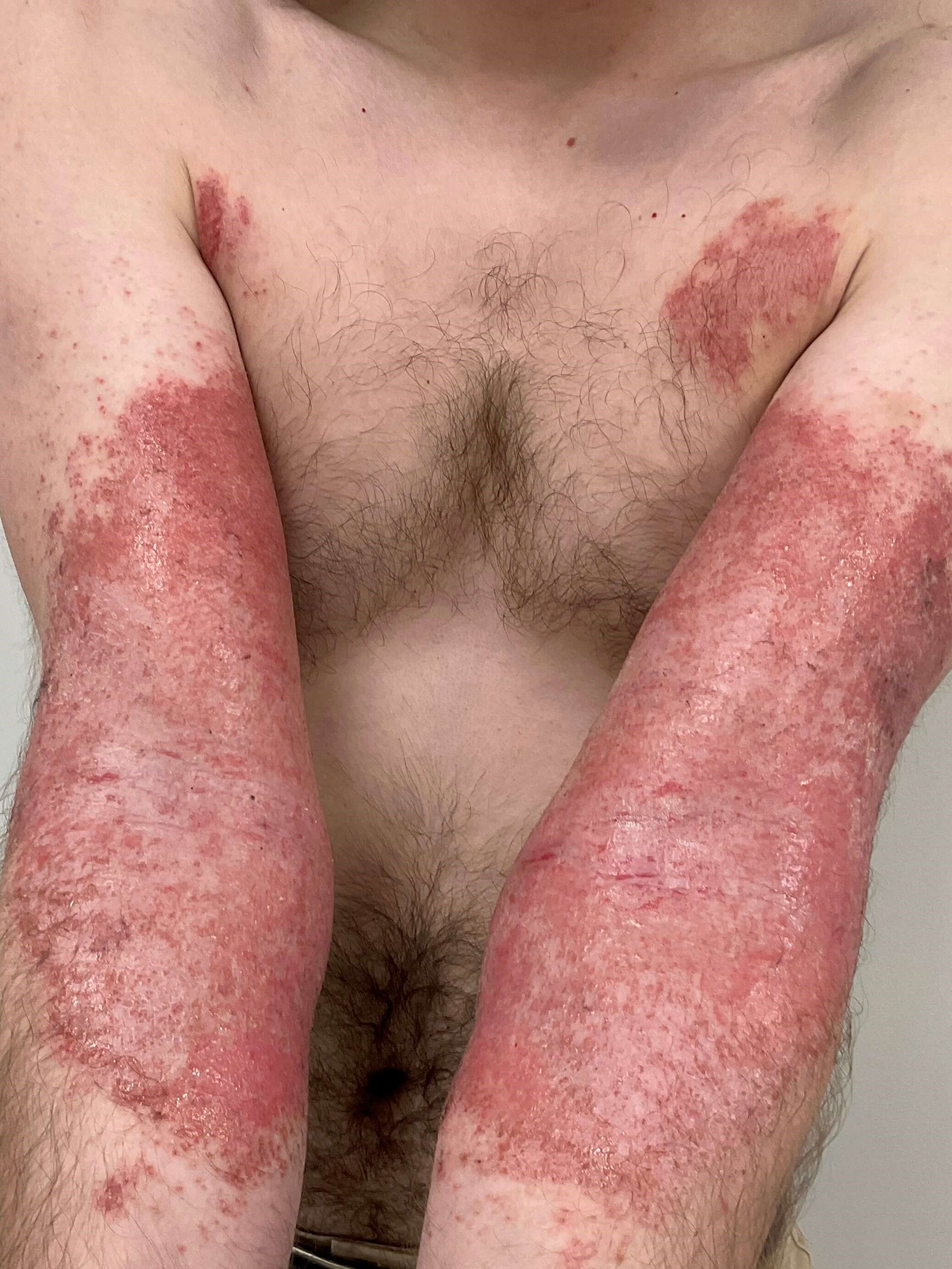
Eczema/Atopic dermatitis
&w=538&do-not-index "Eczema/Atopic dermatitis Before")
&w=&do-not-index "Eczema/Atopic dermatitis Before")
&w=538&do-not-index "Eczema/Atopic dermatitis After")
&w=&do-not-index "Eczema/Atopic dermatitis After")
Eczema/Atopic dermatitis
&w=538&do-not-index "Eczema/Atopic dermatitis Before")
&w=&do-not-index "Eczema/Atopic dermatitis Before")
&w=538&do-not-index "Eczema/Atopic dermatitis After")
&w=&do-not-index "Eczema/Atopic dermatitis After")
Eczema/Atopic dermatitis
&w=538&do-not-index "Eczema/Atopic dermatitis Before")
&w=&do-not-index "Eczema/Atopic dermatitis Before")
&w=538&do-not-index "Eczema/Atopic dermatitis After")
&w=&do-not-index "Eczema/Atopic dermatitis After")
Eczema/Atopic dermatitis
Synopsis
https://www.visualdx.com/visualdx/diagnosis/?moduleId=101&diagnosisId=51378
Atopic dermatitis (eczema) is a chronic, relapsing, pruritic condition that is often associated with allergic rhinitis and/or asthma. Infants and children are most often affected, with 85% of cases appearing in the first year of life and 95% of cases appearing by 5 years. Uncommonly, the condition may persist into, or even arise in, adulthood. Less than 1% of adults are affected by atopic dermatitis. With increased understanding of immunosenescence, atopic dermatitis is increasingly being recognized in the older adult population.In infants, the disease involves primarily the face, scalp, torso, and extensor aspects of extremities. In children and adults, the disease usually involves chiefly the flexural aspects of extremities, but it may be more generalized. In adults, flexural skin may be clear and disease may be focal or widespread. Follicular patterns of atopic dermatitis (ie, follicular eczema) are more common in persons with darker skin phototypes.
Atopic dermatitis may be categorized as follows:
- Acute – erythema, vesicles, bullae, weeping, crusting
- Subacute – scaly plaques, papules, round erosions, crusts
- Chronic eczema – lichenification, scaling, hyper- and hypopigmentation
Intense pruritus (itching) is a hallmark of atopic dermatitis. Scratching leads to lichenification (skin thickening from chronic trauma). Impaired barrier function leads to increased water loss and cutaneous infections. Patients with atopic dermatitis are prone to impetiginization with Staphylococcus aureus. Secondary infections with herpes simplex virus (eczema herpeticum), Coxsackie viruses (eczema coxsackium), or vaccinia virus (eczema vaccinatum) may transpire.
Patients with atopic dermatitis have difficulties in retaining skin moisture and suffer from xerosis (dry skin). Environmental triggers, such as heat, humidity, detergents / soaps, abrasive clothing, chemicals, smoke, and even stress, tend to aggravate the condition. Latex allergy and nickel allergy occur more often in persons with atopic dermatitis. Additionally, patients with atopic dermatitis have been found to be more likely to have positive patch test results to products commonly found in topical treatments, including cocamidopropyl betaine, wool alcohol / lanolin, and tixocortol pivalate. Allergy to eggs, cow's milk, or peanuts is common. There may be a relationship between atopic dermatitis and the development of aspirin-related respiratory disease.
Codes
ICD10CM:L20.9 – Atopic dermatitis, unspecified
SNOMEDCT:
24079001 – Atopic dermatitis
Look For
Scaly, erythematous papules and plaques involving the flexural surfaces, particularly the antecubital fossae and popliteal fossae, face, neck, and extremities in general. In chronic cases, lichenification, scaling, and hyper-, hypo-, and depigmentation may be seen.Follicular eczema presents with scaly follicular-based papules.
Impact of skin color on clinical presentation:
- In darker skin colors, erythema may be subtle and overlooked, including in patients with erythroderma. Gentle pressure to blanch areas of involvement may help reveal subtle erythema upon removal of pressure on skin. Look for the presence of vesicles in acute eczema; scaling, desquamation, hyperpigmentation, erosions, and crusts in subacute eczema; and hyperpigmentation, lichenification, scaling, and desquamation in chronic eczema.
- Pigmentary changes are more prominent in darker skin colors and include postinflammatory hyper-, hypo-, and/or depigmentation after resolution of inflammation. Patchy-to-diffuse hyperpigmentation may also be a sign of active disease (especially in patients with erythroderma in whom hyperpigmentation and superficial desquamation may be the only signs).
- In lighter skin colors, follicular papules of follicular eczema appear skin-colored or erythematous. In darker skin colors, erythema may be subtler, and papules and plaques may appear violaceous or brown.
- Periorbital hyperpigmentation is more overt in patients with darker skin colors.
Associated physical findings include hyperlinear palms and keratosis pilaris (keratotic follicular lesions) on the upper arms, legs, cheeks, and buttocks.
Secondary bacterial infection is a common concomitant finding. Impetiginized plaques may develop thick yellow (honey-colored) crusts. Painful monomorphic crusted erosions may be a sign of a superimposed disseminated HSV infection (Kaposi varicelliform eruption).
Diagnostic Pearls
Make sure to obtain an adequate childhood and family history of allergies, asthma, and skin disease. In general, it is rare for an adult without a personal or family history of atopy to develop atopic dermatitis. Such patients should be referred to dermatology to rule out another entity, specifically mycosis fungoides.In the adult patient, persistent dry skin or persistent eyelid dermatitis may be a clue to atopic dermatitis.
Differential Diagnosis & Pitfalls
- Allergic contact dermatitis
- Irritant contact dermatitis
- Nummular dermatitis (nummular eczema)
- Eczema craquelé
- Tinea corporis
- Psoriasis
- Pityriasis rosea
- Seborrheic dermatitis
- Lichen simplex chronicus
- Ichthyosis vulgaris
- Scabies
- Pityriasis rubra pilaris
- Secondary syphilis
- Glucagonoma syndrome
- Pellagra
- Mycosis fungoides – If an adult patient has persistent "eczema" that is not adequately responding to therapy, this entity should be ruled out with skin biopsies.
Best Tests
A careful history, to include an appropriate temporal course and family history of atopy, coupled with the appropriate clinical appearance, are keys to diagnosis. Serum immunoglobulin E (IgE) level is elevated in 80% of patients, although in routine cases, IgE levels usually are not necessary.Bacterial culture should be sent if lesions appear impetiginized. A Tzanck smear, viral culture, and/or viral polymerase chain reaction (PCR) should be performed if eczema herpeticum is considered.
Skin scrapings for scabies should be performed on any lesion that resembles a burrow.
In a few select cases, the following investigations may help rule out imitators:
- Skin biopsy
- Oral food challenges, radioallergosorbent test (RAST), or patch testing
- HIV test
Management Pearls
Counsel patients on avoiding triggers. Factors that are known to exacerbate atopic dermatitis include stress, inappropriate bathing habits (eg, prolonged, hot showers), infection, irritants (eg, detergents), sweating, and environmental allergens.Appropriate skin care is critical. Gentle nonsoap cleansers should be utilized. The liberal use of bland emollients is essential. These products should be fragrance and dye free.
Patients are prone to bacterial, fungal, or viral superinfections, which can further exacerbate dermatitis flares. Evidence of hemorrhagic crusts (scabs) may be indicative of staphylococcal colonization or viral superinfection.
Sleep disturbance and depression may be seen in this population and should be adequately assessed for.
There has been an association between atopic dermatitis and osteoporotic fractures, although the mechanism is unclear.
Refer chronic, recalcitrant, or severe cases to a dermatologist.
Therapy
Intermittent topical corticosteroids are used to treat disease flares. Class 6-7 topical steroids can be used on the face, and mid- to high-potency preparations can be used on the trunk and extremities. Achieve clearance of dermatitis with the lowest-strength topical steroid that is effective. Atrophy, hypopigmentation, and striae are potential risks of inappropriate use. Ointments without preservatives are preferred. Application to damp skin or under occlusion can enhance penetration.Topical nonsteroidal agents (see below) can be used instead of topical steroids to prevent topical steroid side effects, especially on eyelid skin, the rest of the face, and in folds, or in any chronically active case.
Localized Disease on the Body:
Mid-potency topical corticosteroids (class 3-4) need supervision with scheduled follow-up to observe for steroid atrophy.
- Triamcinolone cream, ointment (0.1%) – Apply twice daily (15, 30, 60, 120, 240 g).
- Mometasone cream, ointment (0.1%) – Apply twice daily (15, 45 g).
- Fluocinolone cream, ointment (0.025%) – Apply twice daily (15, 30, 60 g).
Use low-potency topical steroids on areas of thinner skin on the face and intertriginous areas.
- Desonide or alclometasone ointment twice daily (30 g).
- Tacrolimus ointment (0.03%, 0.1%) twice daily.
- Pimecrolimus cream (1%) twice daily.
- Crisaborole ointment (2%) twice daily; it is a nonsteroidal phosphodiesterase 4 inhibitor for use in children (aged 2 years or older) and adults with mild-to-moderate atopic dermatitis. Early data show rapid relief of pruritus and clinical improvement in patients.
- Ruxolitinib cream (1.5%) twice daily; it is a topical selective JAK1 and 2 inhibitor approved for patients 12 years and older with mild-to-moderate atopic dermatitis inadequately controlled by (or patient is unable to use) other topical therapies. Topical ruxolitinib should not be combined with other biologic or immunosuppressive therapies, including other JAK inhibitors. As with other JAK inhibitors, ruxolitinib carries a black box warning that should be discussed as warranted.
Extensive Disease:
Detailed review and discussion of treatment of extensive disease has been studied. Controversy still exists on the best approach to these therapies, and the guidelines should be reviewed when considering the treatment of individual patients.
Dupilumab, a human monoclonal antibody inhibiting interleukin (IL)-4 and IL-13, is approved for adults and children aged 6 months or older with moderate-to-severe atopic dermatitis inadequately controlled by or unable to use topical steroid treatment. It is administered by subcutaneous injection.
Systemic therapies include nonsedating antihistamines and systemic antibiotics for definitely infected disease. Systemic steroids should be avoided due to the risk of rebound flares. Their use should strictly be reserved for acute and severe flares in conjunction with other treatment modalities.
Cyclosporine, azathioprine, tacrolimus, and mycophenolate mofetil have all been used to treat extensive, resistant disease with varying degrees of success. Phototherapy – including UVB, psoralen plus UVA (PUVA), and narrowband UVB – has also been used successfully in many patients. Narrowband UVB when used must be modified for the skin type of the individual.
Abrocitinib is US Food and Drug Administration (FDA) approved for the treatment of moderate-to-severe atopic dermatitis in adults who are 18 years or older and in whom disease is not controlled with other systemic agents. The recommended dose is 100 mg daily, and 200 mg daily if there is lack of response to the 100-mg dose. Lower dosing recommendations (50 mg/day) are also put forward for patients with moderate renal failure, and for patients receiving medications that inhibit cytochrome P450 (CYP) 2C19 inhibitors. Upadacitinib, another selective JAK1 inhibitor, has also been FDA approved for moderate-to-severe atopic dermatitis in adults and children aged 12 years and older whose disease did not respond to prior oral and injectable therapies. The starting dose is 15 mg daily for patients weighing 40 kg or more, and this can be increased to 30 mg daily in qualified patients 65 and younger if needed. Two phase 3 trials of baricitinib showed significant improvement in moderate-to-severe atopic dermatitis for 2-mg and 4-mg doses compared with placebo.
As with other oral JAK inhibitors used to treat chronic inflammatory conditions, an FDA black box warning has been issued for baricitinib and upadacitinib due to increased risk of serious infections, heart-related events, stroke, heart attack, blood clots, cancer, and death. Abrocitinib is undergoing further safety evaluation by the FDA. This warning was issued based on postmarketing data in patients with rheumatoid arthritis, and long-term safety data in patients with inflammatory skin disease are not yet available. These warnings, as with all medication side effects, should be discussed with patients to determine if potential benefits outweigh potential risks of therapy.
Tralokinumab is a monoclonal antibody against IL-13. It is approved for use in adults with moderate-to-severe atopic dermatitis not responsive to topical therapies or in whom other therapies are not tolerated. It is administered subcutaneously in an initial dose of 600 mg, then 300 mg every other week. At 16 weeks of treatment, a maintenance dose of 300 mg every 4 weeks may be considered.
These treatments are best administered by a dermatologist familiar with their use.
Pruritus:
Antihistamine therapy may serve as an adjunct for the management of pruritus. Antihistamines are not recommended as first-line therapy but may be useful if the patient has sleep disruption, significant dermatographism, or other seasonal atopy symptoms. Consider one of the following antihistamines.
- Diphenhydramine hydrochloride (25, 50 mg tablets or capsules): 25-50 mg nightly or every 6 hours as needed.
- Hydroxyzine (10, 25 mg tablets): 12.5-25 mg, every 6 hours as needed.
- Cetirizine hydrochloride (5, 10 mg tablets): 5-10 mg/day.
- Loratadine (10 mg tablets and RediTabs): 10 mg tablet or RediTab once daily.
Infection:
Antibiotic therapy is beneficial when there is evidence of impetiginization (honey-colored crust, denuded skin, or oozing). Treat with a 10-day course of oral antibiotics to cover S aureus infection. As methicillin-resistant S aureus (MRSA) becomes more prevalent in the community, therapy should be appropriately tailored. Mupirocin 2% topical ointment can be used as a topical adjunct. Bleach baths (1/4 to 1/2 cup household bleach diluted in a full bathtub of water) 1-3 times weekly may be useful in patients with multiple open areas (excoriations) or a history of multiple superinfections.
Secondary infection due to herpes simplex virus (eczema herpeticum) should be treated with antiviral therapy (acyclovir or prodrug forms).
Location
* All information subject to change. Images may contain models. Individual results are not guaranteed and may vary.